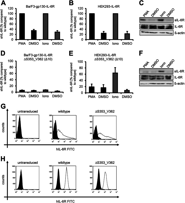
FIGURE 1.
ADAM17 and ADAM10 use different cleavage sites of the human IL-6R. A, Ba/F3-gp130-hIL-6R cells were treated for 2 h with PMA (100 nm) or for 1 h with ionomycin (Iono, 1 μm). Soluble IL-6R was measured by ELISA. The amount of soluble cytokine receptor without stimulation was considered as constitutive shedding and set to 1. Based on this, the increase of soluble receptors was calculated. B, HEK293 cells were transfected with an expression plasmid encoding wild-type human IL-6R. Cells were treated as described in panel A, sIL-6R was measured by ELISA, and values were calculated accordingly. C, transiently transfected HEK293 cells were treated as described under panel A. To determine the soluble cytokine receptors via Western blotting, they were precipitated from conditioned media with concanavalin A-covered Sepharose beads and visualized with 4-11 antibody (IL-6R). Cells were lysed after stimulation, and lysates were subsequently analyzed via Western blotting, whereas β-actin served as loading control. D, Ba/F3-gp130-IL-6RΔS353_V362 cells were treated as described in panel A. E and F, HEK293 cells were transfected with an expression plasmid encoding hIL-6RΔS353_V362. The experiment was performed as described in panels A and B. ELISA data are the mean (±S.D.) from three independent experiments, and Western blotting shows one representative experiment. G, cell surface expression of wild-type IL-6R and IL-6RΔS353_V362 on transiently transfected HEK293 cells was determined via flow cytometry as described under “Experimental Procedures.” H, cell surface expression on stably transduced Ba/F3-gp130-hIL-6R and Ba/F3-gp130-IL-6RΔS353_V362 cells was determined via flow cytometry as described under “Experimental Procedures.” One representative experiment of three performed is shown. The expressed IL-6R variant is given above the respective FACS plot.

