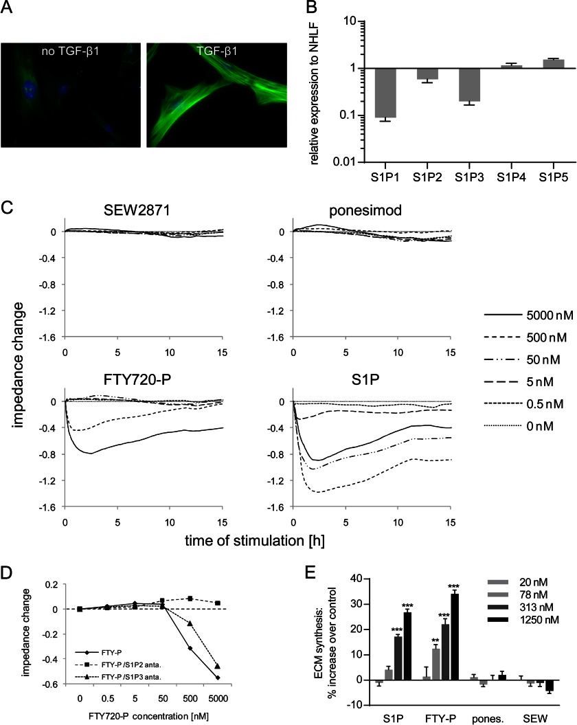
FIGURE 7.
Analysis of S1PR mRNA expression, signaling, and ECM induction in NHLF-derived myofibroblasts. A, NHLF were treated with or without 1 ng/ml TGF-β1 for 72 h, reseeded, and starved for 24 h before immunofluorescent staining was performed. Green, α-smooth muscle actin; blue, nuclei. B, expression changes of S1PR mRNA in myofibroblasts compared with nontransformed NHLF. The data show the means ± S.E. of three independent experiments. C, myofibroblasts were stimulated with SEW2871, ponesimod, FTY720-P, or S1P (0.5–5000 nm), and impedance responses were monitored for 15 h. D, concentration response curves for FTY720-P in the absence or presence of S1P2R or S1P3R antagonists generated from the impedance values at 90 min. The data in C and D show representative experiments (n = 2–3). E, myofibroblasts were stimulated with S1PR agonists (20–1250 nm), and ECM synthesis was measured after 24 h with the [3H]proline incorporation assay. The data represent the means ± S.E. of three independent experiments. *, p < 0.05; ***, p < 0.001, one-way analysis of variance, Dunnett's post test.

