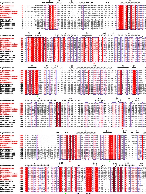
FIGURE 4.
Multiple-sequence alignment of BglA-2 and homologs. The 6-phospho-β-glucosidases and 6-phospho-β-galactosidases are labeled with red and black titles, respectively. Two conserved catalytic glutamate residues and the subsite −1 residue Trp415 are depicted by green triangles. The subsite +1 residues and the phosphate-binding residues are marked with red and black triangles, respectively. The tryptophan residue discriminating 6-phospho-β-galactosidase from 6-phospho-β-glucosidase in GH-1 family is indicated by a blue triangle.

