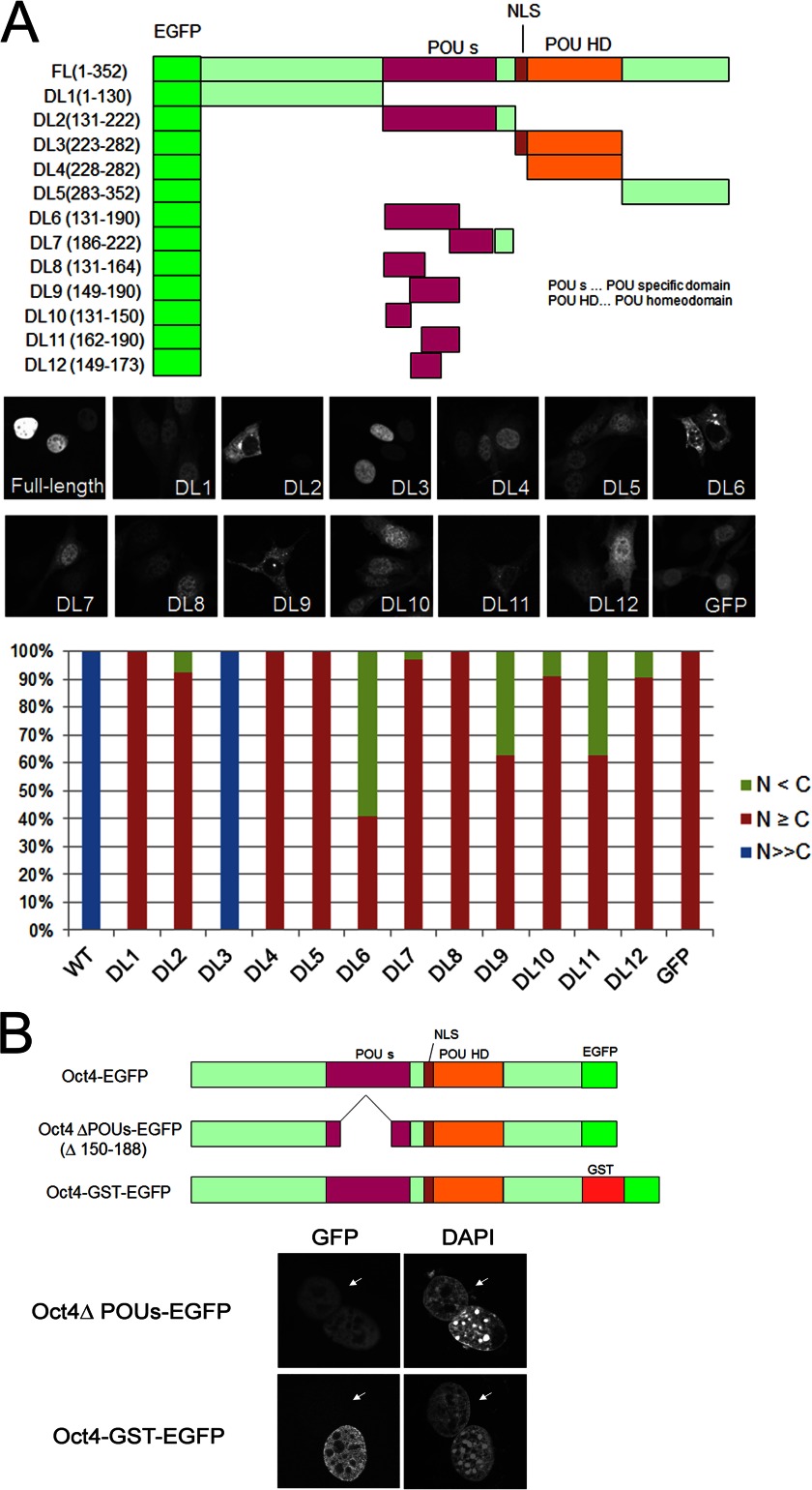
FIGURE 2.
The POU-specific domain is not essential for Oct4 shuttling activity. A, shown is a schematic diagram and cellular localization of various Oct4 deletion mutants N-terminally tagged with EGFP. The regions corresponding to POUS, NLS, and POUHD are indicated. NIH3T3 cells were transfected with the constructs, and cells were fixed, permeabilized, and counterstained with DAPI at 24 h post-transfection. Subcellular localization of EGFP-Oct4 and its mutants was classified into three categories: nuclear (blue), nuclear dominant (red), and cytoplasmic dominant (green). B, shown is a schematic representation of the Oct4 ΔPOUs-EGFP and Oct4-GST-EGFP fusion proteins and heterokaryon assay results. NIH3T3 cells were infected with retroviruses expressing Oct4 ΔPOU-EGFP or Oct4-GST-EGFP, and heterokaryon assays were performed as described in Fig. 1. Arrows indicate the nuclei of HeLa cells.

