FIGURE 3.
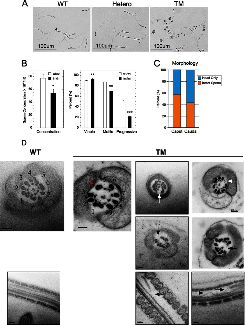
Abnormal sperm in Stamptm/tm mice. A, morphology of Stamp+/+ (WT), Stamp+/tm (Hetero), and Stamptm/tm (TM) male mice (12 weeks old) was determined by light microscopy. Sperm was isolated from epididymis and stained with 0.5% trypan blue. B, epididymal spermatozoa were analyzed by computer-assisted sperm analysis to determine concentration, viability, and motility. Viability was defined by the ability to exclude fluorescent Viadent; motility was defined by the ratio of the motile to the total sperm concentration; and progressive motility was defined by sperm moving faster than the minimum path velocity (VAP). The data are expressed as the means ± S.E. of eight independently obtained biological samples of >99% C57/Bl6 mice (each >500 sperm). *, p < 0.05; **, p < 0.005; ***, p < 0.0005. C, sperm isolated from caput or cauda epididymides of Stamptm/tm mice were imaged by light microscopy. The percentage of disrupted sperm was determined from the number of detached sperm heads and intact sperm in three independent (>500 sperm) biological samples. Using a Student's t test, there was no statistically significant difference between Stamptm/tm sperm isolated from the caput and cauda epididymis. D, disrupted axoneme in Stamptm/tm sperm tails. Electron microscopy detected the canonical arrangement of nine peripheral microtubule doublets, and the larger associated, more peripheral outer dense bodies or fibers, surrounding a central doublet (9 + 2) that were intact in transverse and longitudinal sections in 100% of normal sperm tails. The top left panel is from WT sperm, with the numbering of the nine microtubule doublets. In Stamptm/tm sperm, doublet 4 is missing from the 9 + 2 formation in virtually all of the axonema (see top left panel of TM pictures for numbering, with the red number 4 indicating the position of missing double 4 and white arrows for other examples). Severe disruption of microtubule polymers (arrows in bottom row) was observed in transverse sections. Occasionally, more severe defects were observed (middle row). The more peripheral dense fibers were generally indistinguishable from normal controls except that the outer dense fiber paired with doublet 4 usually disappears with doublet 4. The samples were independently obtained from three biological samples. Scale bars, 0.2 μm.

