FIGURE 6.
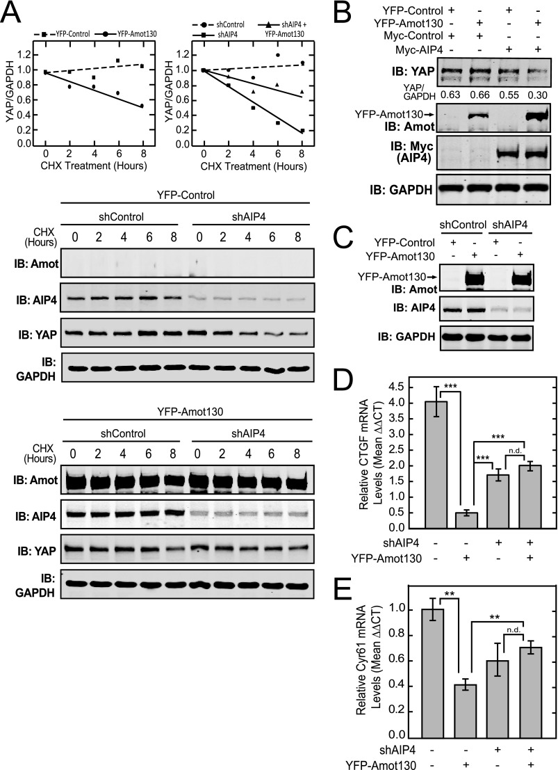
Amot130 and AIP4 interdependently inhibit YAP. A, the levels of YFP-tagged Amot130 as well as endogenous AIP4, YAP, and GAPDH were detected in lysates from MDA-MB-468 cells stably expressing control or AIP4 shRNA in combination with YFP-tagged Amot130 or control vector and treated for the indicated times with vehicle (dimethyl sulfoxide) or 200 μg/μl of cycloheximide (CHX). The ratio of pixel intensities with linear regression analysis was plotted from the immunoblots, where the YAP/GAPDH levels in cells expressing YFP-control (■) or YFP-Amot130 (●) are depicted (left graph) and the YAP/GAPDH levels in cells expressing shControl (●), shAIP4 (■), or YFP-tagged (▴) Amot130 and shAIP4 are shown (right graph). B, the levels of endogenous YAP were detected by immunoblot (IB) analysis of lysates from HEK 293T cells expressing combinations of YFP-tagged Amot130 and Myc-tagged AIP4. The ratio of pixel intensities of endogenous YAP over GAPDH bands are below the blot. C, MDA-MB-468 cells stably expressing YFP-tagged Amot130 or control vector in combination with control or AIP4 shRNA. The mRNA transcript levels from cells described in C of CTGF (D) or Cyr61 (E) were measured by real-time quantitative PCR. Error bars represent ± S.D. ***, p < 0.00001; **, p < 0.01; n.d., no statistical difference.

