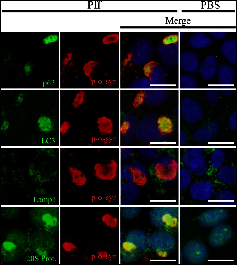
FIGURE 1.
Co-localization of protein degradation pathway components with α-syn aggregates. IF images of HEK293 α-syn cells 24 h after Pff transduction, stained with p-α-syn antibody (red), DAPI (blue), or antibodies to p62, LC3, Lamp1, and 20 S proteasome (green). Dispersed and punctate cytoplasmic staining of p62 and LC3 was observed in PBS-td cells. In Pff-td cells, p-α-syn aggregates showed strong co-localization with p62 and LC3. Similarly, there was diffuse staining of the 20 S proteasome in the PBS-td cells, whereas Pff-td cells had cytoplasmic 20 S proteasome staining that largely co-localized with α-syn aggregates. In contrast, Lamp1-positive vesicles were observed in both PBS-td and Pff-td cells, and these did not typically co-localize with p-α-syn aggregates. Scale bars, 20 μm.

