FIGURE 12.

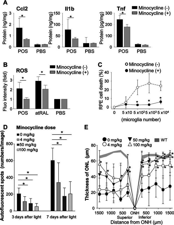
Minocycline inhibits activation of microglia in vitro and attenuates light-induced retinal degeneration in vivo. Retinal microglia (5 × 10³/well in 96-well plates) isolated from 2-week-old C57BL/6 WT mice were incubated at 37 °C with or without 6 μg/100 μl of POS from 4-week-old WT mice. Cultured cells were divided into two groups without or with minocycline (30 μg/ml). A, production of Ccl2, Il1β, and Tnf was quantified by ELISA using culture supernatants of microglia cells with or without 24 h stimulation by POS. Minocycline was administrated as noted above. Error bars indicate mean ± S.D. (n = 6–9). * indicates p < 0.05. B, generation of ROS was examined by the ROS probe, DCF-DA. After 24 h incubation, cells were washed with PBS twice and 0.25 μm DCF-DA was co-incubated for 30 min. Error bars indicate mean ± S.D. (n = 3). * indicates p < 0.05. C, the influence of minocycline was tested using the RPE cell death system described in the legend to Fig. 6B. Minocycline administration prevented RPE cell death caused by microglia activation. Error bars indicate mean ± S.D. (n = 3). * indicates p < 0.05. D and E, the therapeutic effect of minocycline in vivo was tested in light-induced retinal degeneration model. Minocycline was administrated as described under ”Experimental Procedures.“ Minocycline administration attenuated AF spot accumulation (D) observed by using SLO, and retinal degeneration (E) represented by ONL thickness measured by OCT in a dose-dependent manner.
