FIGURE 6.

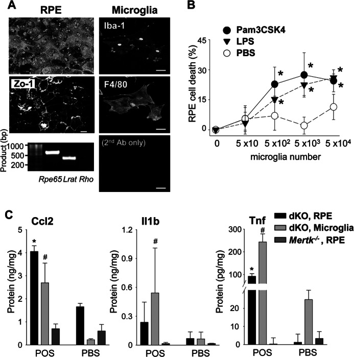
Primary cultured RPE and microglial cells produce Ccl2, Il1b, and Tnf in response to photoreceptor proteins. A, primary RPE and retinal microglial cells were isolated from 2-week-old Abca4−/−Rdh8−/− mice. Morphology of RPE cells after 7 days in culture was captured with bright-field microscopy (left upper), and immunohistochemistry with anti-Zo-1 Ab was carried out (left middle). RT-PCR showed RPE-specific Rpe65 and Lrat amplification at day 14 in the primary RPE culture (left bottom) without photoreceptor-specific rhodopsin (Rho) amplification. Cultured microglia at 7 days displayed microglia- and macrophage-specific staining with anti-Iba-1 and anti-F4/80 Abs (right). Bars indicate 10 μm. B, primary RPE cells from 2-week-old Abca4−/−Rdh8−/− mice (5 × 10³) were co-incubated with different numbers of microglia cultured for 7 days with or without 1 μm Pam3CSK4 or 1 μm LPS for 24 h, and the activity of LDH released from dead cells was measured. Error bars indicate mean ± S.D. (n = 3). * indicates p < 0.05 versus PBS. C, production of Ccl2, Il1b, and Tnf was quantified by ELISA with culture supernatants of primary cultured RPE or microglia from 2-week-old Abca4−/−Rdh8−/− (dKO) or Mertk−/− mice after co-incubation with or without 6 μg/100 μl of mouse POS for 24 h. Error bars indicate mean ± S.D. (n = 9). * and # indicate p < 0.05 versus PBS.
