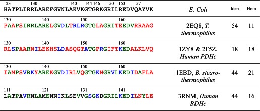
TABLE 6.
Sequences of the E3-binding domains
Sequences of known crystal structures of E3-binding domains from different sources are compared with that of the E3-binding domain from the dihydrolipoamide succinyltransferase core from the pyruvate dehydrogenase multienzyme complex of E. coli. Residues at equivalent locations identical to those in E. coli are shown in green, homologous residues in blue, and other residues in red. The last two columns indicate the percentage of residues identical with (Iden) or homologous to (Hom) those in E. coli.