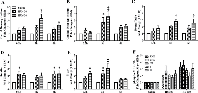
Fig. 3.
Intensity means of phospho-MEK-3/6 in different regions of GD9 embryos. The quantification of phospho-MEK-3/6 immunoreactivity by intensity mean in GD9 embryos exposed to saline or HU at 400 or 600mg/kg in different regions is presented in the RNE (A), CNE (B), NT (C), somite (D), and heart (E). Values were normalized to the corresponding saline control and expressed as fold changes. n = 5. Asterisks, daggers, and double daggers denote a significant difference from saline control at the same time point (*p < 0.05; †p < 0.01; ‡p < 0.001, Dunnett’s test). (F) Phospho-MEK-3/6 staining at 3h post-HU treatment. There were no significant regional differences in phospho-MEK-3/6 staining within the embryo.

