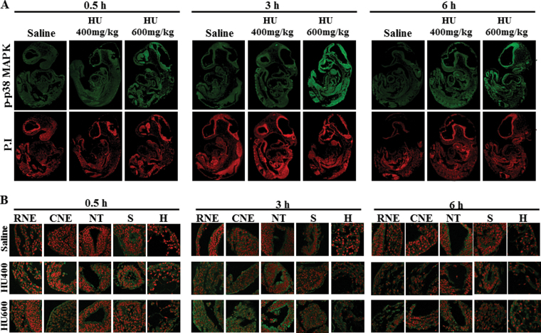
Fig. 5.
The localization of phospho-p38 MAPK in GD9 embryos exposed to HU. Timed-pregnant female mice received saline or HU at 400 or 600mg/kg on GD9. Embryos were processed for immunofluorescence staining with an antibody against phospho-p38 MAPK (in green) and counterstained with propidium iodide (in red). (A) Whole embryo views of phospho-p38 MAPK immunoreactivity at 0.5, 3, and 6h posttreatment. (B) 60× magnification views of the selected five regions in an embryo: RNE, CNE, NT, somite (S), and heart (H).

