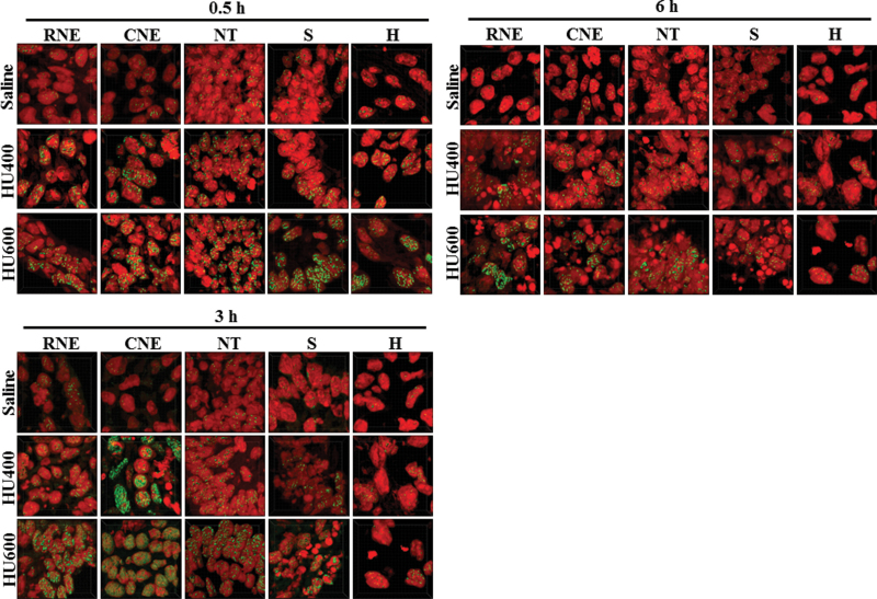
Fig. 8.
γH2AX staining in GD9 embryos exposed to HU. The detection of DNA DSBs with γH2AX in saline and HU-exposed embryos in different regions. Immunodetection of γH2AX is in green and nuclear propidium iodide in red. Images display quantification regions (50 × 50 × 14 µm), which were randomly selected from each structure taken at 63× magnification (shown in Supplementary fig. S4).

