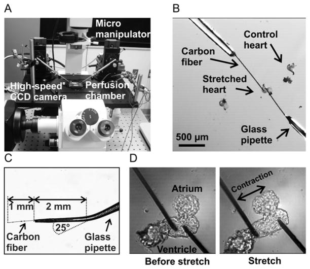
Fig. 1. Experimental system for the application of controlled stretch to zebrafish embryo hearts.
The zebrafish heart stretch system was designed using a conventional inverted fluorescence microscope with attached micromanipulators (panel A). A high-speed CCD camera was used for measuring action potentials and conduction velocities. Two carbon fibers were attached to the ventricle of an isolated zebrafish embryo heart (panel B). The terminal 2 mm of a glass pipette was bent by 25° and a 1 mm long section of carbon fiber was glued into each pipette using a cyanoacrylate-based adhesive (panel C). Zebrafish hearts were stretched from the force-free position (panel D, left) to the stretched position (panel D, right) using the micromanipulators.

