
Fig. 4. Time course study to determine the optimal time point for stretch experiments in the developing zebrafish embryo heart.
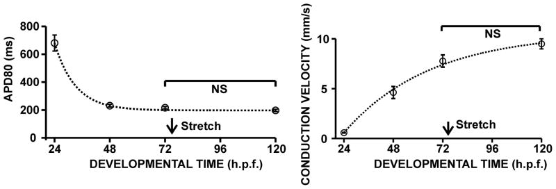
Ventricular action potential durations (APD80, left) and ventricular conduction velocities (right) were calculated from hearts isolated in 24 hour intervals, between 24 and 120 h.p.f. to evaluate the influence of development on electrical activity during the time of stretch. The time point of 72 h.p.f. was chosen for all subsequent stretch experiments and is indicated by the arrow.