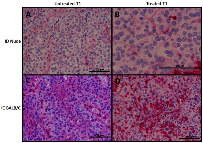
Figure 7. CD3+ Immunohistochemsistry of primary (T1) tumors.
CD3+ staining, indicative for T-cell presence, performed for (A,C) untreated and (B,D) treated initial T1 tumors between (A,B) ID nude and (C,D) IC BALB/c mice. There is no notable difference observed in CD3+ infiltration for ID nude mice between (A) untreated and (B) treated tumors. For the IC BALB/c mice, a robust increase in CD3+ (T-cell) infiltration is observed in some treated tumors (D) relative to untreated T1 controls (C). Increased T-cell presence in treated T1 IC mice was also more robust than for both groups for nude mice (A,B). All scale bars 200 µm. Panels (A,C,D) 200x, panel (B) 400x magnification.

