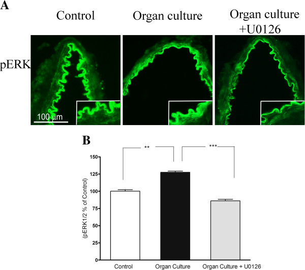
Figure 6.

A. Sections from the human cerebral artery showing pERK immunoreactivity in the smooth muscle cell layer. There are increased expressions of the ERK1/2 protein levels in the cultured arteries compared to the control segments. Treatment with U0126 prevented the increased protein expression in the smooth muscle cells. B. Expression of pERK1/2 protein levels in human cerebral arteries incubated for 48 h with or without the MEK1/2 inhibitor U0126 (5 μM) and control human arteries. Data are expressed as percentage of fresh and given as mean ± s.e.m. *P<0.05, ** P<0.01.
