Figure 2.
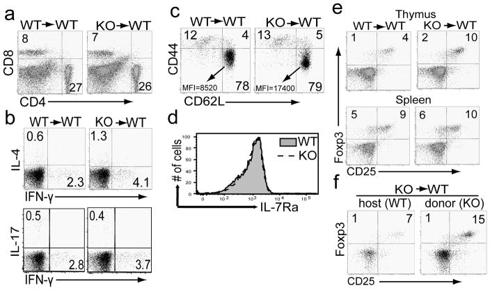
Sin1 deficiency increases the number of thymic natural regulatory T cells
a) Splenocytes from Sin1+/+ (WT) or Sin1−/− (KO) chimeric mice were analyzed for percentages of CD4 and CD8 T cells. The FACS plots shown are pre-gated on CD45.2+ donor cells.
b) Splenocytes from Sin1+/+ (WT) or Sin1−/− (KO) chimeric mice were stimulated with PMA and ionomycin for 4 hours in the presence of Golgi-Stop and assayed for IFN-γ, IL-17A, and IL-4 expression by flow cytometry. The FACS plots shown are pre-gated on donor CD4+CD8− CD45.2+ cells.
c) The expression of CD62L and CD44 was measured on splenic CD4+ T cells from Sin1+/+ or Sin1−/− chimeric mice by flow cytometry. The mean fluorescent intensity (MFI) of CD62L in the CD44lowCD62Lhi gate are as follows: Sin1+/+ MFI=8520 and Sin1−/− MFI=17400. The FACS plots shown are pre-gated on donor CD4+CD8− CD45.2+ cells.
d) The expression of IL-7R (CD127) on splenic donor CD4+CD45.2+ T cells from Sin1+/+ or Sin1−/− chimeric mice was determined by flow cytometry.
e) The thymic (upper panel) and splenic (lower panel) donor CD4+Foxp3+CD45.2+ T cells in Sin1+/+ or Sin1−/− chimeric mice were determined by flow cytometry.
f) Chimeric mice containing host WT and Sin1−/− (KO) T cells were generated by transferring Sin1−/− fetal liver cells into sub-lethally irradiated WT (CD45.1+) hosts. The percent of thymic WT CD4+Foxp3+ T cells (CD45.1+) and donor Sin1−/− CD4+Foxp3+ T cells (CD45.2+) in a chimeric mouse was determined by flow cytometry.
Numbers in the plots indicate percentages of the gated populations. All flow cytometry plots shown are representative of Sin1+/+ (n=3) and Sin1−/− (n=4) fetal liver chimeric mice.

