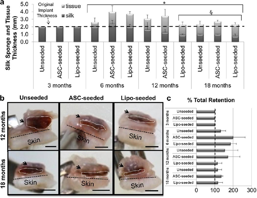
Fig. 2.
Tissue regeneration occurs with silk sponge degradation. (a) The silk sponge volume was calculated by measuring the silk sponge thickness and diameter upon explantation. The volume was maintained, i.e. no biomaterial degradation, through 6 months. At 12 and 18 months, the seeded groups degraded more quickly than the unseeded group. Tissue thickness had significantly increased at 6, 12 and 18 months when compared to 3 months (* indicates p < 0.0001). Sponge size had significantly decreased at 12 months for the lipo-seeded group, and at 18 months for all groups (§ indicates p < 0.05). (b) Cross-sections of 12 (top) and 18 (bottom) month explants are shown for unseeded (left), ASC-seeded (middle) and lipo-seeded (right) groups. The region in the white dotted line outlines the silk sponge, while the black dotted line demarcates the skin from the subcutaneous tissue. The arrows point to regions of subcutaneous fat. The subcutaneous fat formation was greatest in the lipo-seeded group (right column) and least in the unseeded group (left column). Scale bar – 2 mm. (c) Approximated thickness of the newly formed tissue was determined by measuring the thickness of the fat and subtracting the thickness of the silk sponge by image analysis in ImageJ. There were no statistically significant changes in total % retention.

