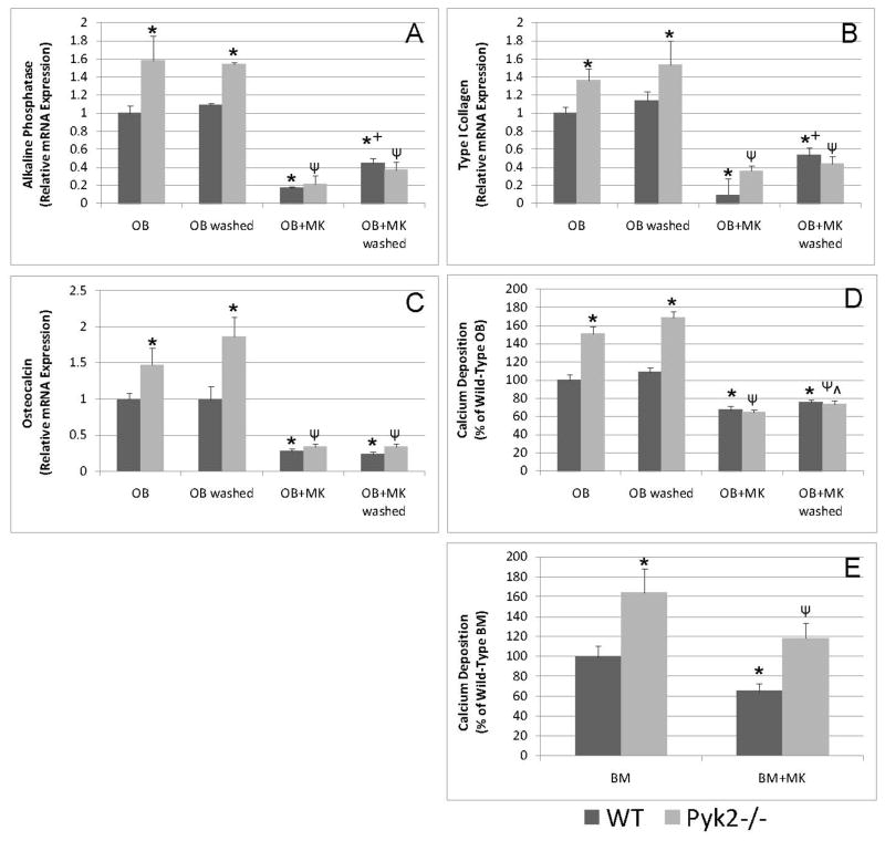
Figure 7.
Real-time quantitative PCR and calcium deposition analyses of OB or BM cells cultured under osteogenic conditions in the presence or absence of MKs for 14 days. A–D) WT and Pyk2−/− OBs were cultured alone or in the presence of MKs for 14 days or for the first 4 hrs of culture and then MKs were removed by washing. OB differentiation as determined by mRNA expression and mineralization was higher in Pyk2−/− OBs cultured alone as compared to that observed in WT OBs cultured alone. Additionally, co-culture of OBs with MKs resulted in a dramatic reduction in OB differentiation irrespective of MK culture duration and in the presence and absence of OB Pyk2 expression. E) To test the effect of MKs on osteogenic progenitors BM cells from WT and Pyk2−/− mice were cultured alone or in the presence of MKs and mineralization assessed on day 14. Pyk2−/− BM cultures were more mineralized than were WT cultures. MKs were able to suppress mineralization in both WT and Pyk2−/− BM cultures. n=3–4/experimental group, representative data are shown from 2 (E&F) or 3 (A–D) experiments conducted. *Indicates statistically significant differences (p<0.05) compared to WT OB or WT BM. ΨIndicates statistically significant difference (p<0.05) compared to Pyk2−/− OB or Pyk2−/− BM. +Indicates statistically significant difference (p<0.05) compared to WT OB+MK. ^Indicates statistically significant difference (p<0.05) compared to Pyk2−/− OB+MK.

