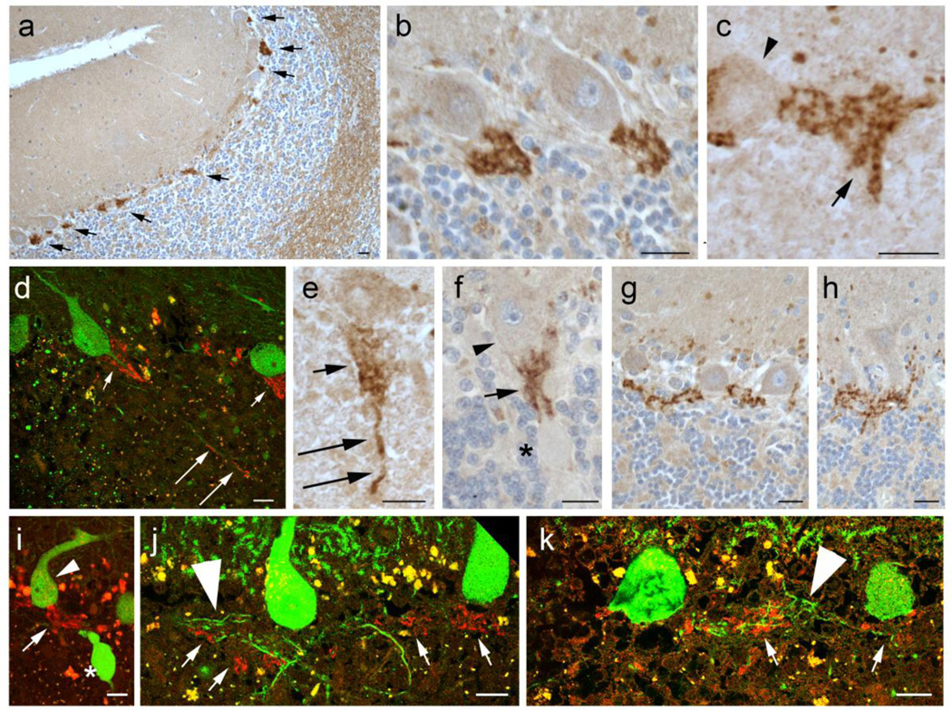
Fig. 2.
Lingo-1 expression in the cerebellar cortex. Immunohistochemistry with Lingo-1 antibody (a–c, e–h) and dual immunofluorescence with anti-Lingo-1 (Alexa 594, red) and anti-calbindinD28k (Alexa 488, green)(d, i–k). Lingo-1 is enriched in a plexus of processes along the PC axon initial segment (AIS) (arrows in a), along with some labeling in white matter along axon profiles and weaker labeling of PC soma and molecular layer (a). The Lingo-1 labeled plexus is often cone-shaped (b–d), and associated with rounded punctate profiles (arrow in c). Lingo-1 labeled processes are occasionally seen along more distal segments of the PC axon (long arrows in d, e). The Lingo-1 labeled plexus surrounds PC axons proximal to axonal torpedoes (asterisk in f, i) and may form complex linear extensions between PC soma (g, h). Lingo-1 labeled plexus may appear in proximity to PC recurrent collaterals (large arrowheads, j,k), but do not consistently colocalize. Small arrowheads: PC bodies; large arrowheads: PC recurrent collateral processes; arrows: Lingo-1 labeled processes; asterisk: torpedo. Scale bar: 20µm.

