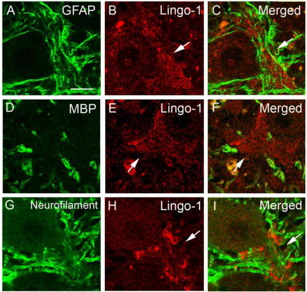
Fig. 3.
Lingo-1 positive plexus did not colocalize with glial markers or neurofilament protein. Dual immunofluorescence labeling of ET cerebellar cortex with anti-GFAP (a), anti-MBP (d), or anti-phosphorylated-neurofilament antibody (g) (Alexa 488, green; a, d, g) and anti-Lingo-1 antibody (b, e, h) (Alexa 594, red). Merged images are shown (c, f, i). Arrow: Lingo-1 positive processes. Scale bar: 25µm

