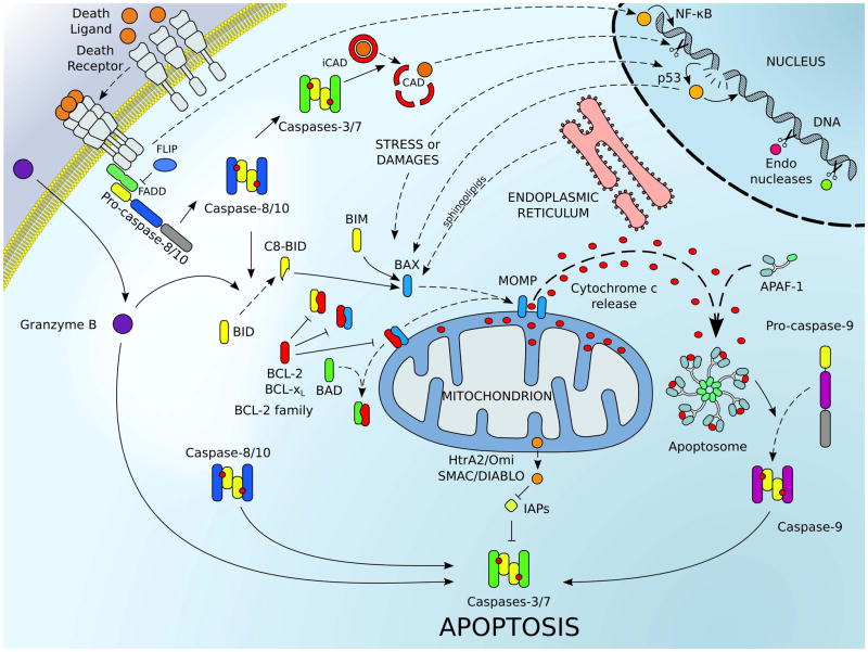
Figure 1.
The major signaling pathways leading to cellular apoptosis. The extrinsic pathway of apoptosis (upper left corner) is engaged by plasma membrane associated death receptors belonging to the TNF-R (tumor necrosis factor receptor) superfamily (e.g., TNF-R1/2, CD95/FAS). Upon engagement of these receptors by their respective ligands (e.g., TNF-α, FASL), conformation changes within the trimerized receptor/ligand complexes recruits adaptor proteins (e.g., FADD) and caspase-8 (and/or caspase-10 in human; represented in blue) to assemble a death inducing signaling complex, referred to as the DISC. Assembly of the DISC promotes caspase-8 activation; cleavage and activation of executioner caspases-3, -6 or -7 (represented in green), and cell death.[165] The intrinsic pathway (also called mitochondrial pathway of apoptosis) responds to cellular stresses like DNA damage (through p53), viral infection, protein misfolding, and oxidative-stress. These signals converge to activate the pro-apoptotic proteins of the BCL-2 family. Pro-apoptotic effectors (e.g., BAX, in blue) are able to target mitochondria and induce mitochondrial outer membrane permeabilization (MOMP); in which numerous pro-apoptotic proteins of the intermembrane space (e.g., cytochrome c, the second mitochondrial-derived activator of caspases SMAC/DIABLO, and HtrA2/Omi) are released into the cytosol. Direct activator BH3-only proteins (e.g., BID and BIM, in yellow) and other signals (e.g., p53 or sphingolipids from the ER) facilitate the activation of BAX and BAK. Sensitizers/de-repressors (e.g., BAD and Noxa, in green) interact with the anti-apoptotic members (e.g., BCL-2 and BCL-xL, in red) to lower the cell death threshold. Once in the cytosol, cytochrome c interacts with the adaptor protein apoptotic protease activating factor 1 (APAF-1) to form the apoptosome, which triggers the recruitment and the activation of the caspase-9 (represented in purple). Inhibitors of apoptosis (IAPs) inhibit caspase activation, and SMAC/DIABLO relieves this inhibition. Once initiator caspases (e.g., caspases-8 and -9) are activated, they trigger downstream activation of effector caspases-3 and -7, which cleave numerous cellular substrates including the inhibitor to the caspase activated DNAse (iCAD). Additionally, granzyme B, which is released by cytotoxic T cells, can also directly trigger effector caspase activation to promote cell death. The extrinsic pathway can also engage the intrinsic pathway via caspase-8-mediated cleavage of BID (in yellow) to amplify pro-apoptotic signaling.

