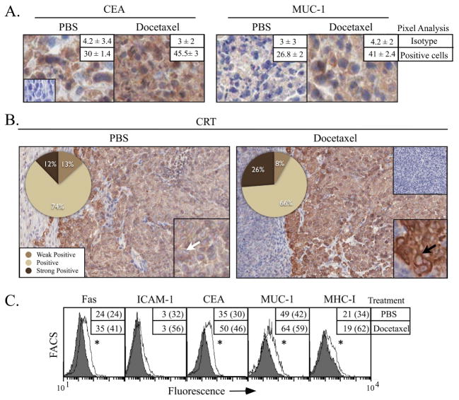
Figure 3.
In vivo treatment with docetaxel modulates tumor phenotype. Nude mice bearing LNCaP xenografts were treated with docetaxel or vehicle (PBS). One week later, tumors were surgically removed, stained, and evaluated by immunohistochemistry for expression of the tumor antigens CEA or MUC-1 (A, 40X, inset: isotype control), or CRT (B, 20X, inset 40X). Numbers indicate percentage of positive cells as determined by pixel analysis (n = 2 mice/treatment group). Arrows indicate CRT membrane staining. (C) Flow cytometry of tumors treated with docetaxel (open histograms) or PBS (shaded histograms). Numbers indicate percentage of positive cells. Numbers in parentheses denote MFI. * = statistical significance relative to untreated cells.

