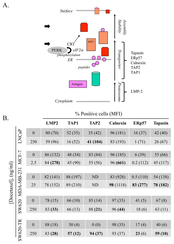
Figure 5.
Tumor cells modulate components of the APM chain after treatment with docetaxel. (A) Schematic of antigen-processing components from cytoplasm to cell surface. Arrows indicate functional checkpoints for CRT and PERK. (B) Human tumor cells were treated in vitro for 72 h with 2.5–250 ng/mL of docetaxel or left untreated, then analyzed for key intracellular components of the APM chain by flow cytometry. Numbers indicate percentage of positive cells. Numbers in parentheses denote MFI. Bold type indicates marked upregulation (≥ 10% increase in percent of cells or 30% increase in MFI not observed in isotype control vs. untreated cells).

