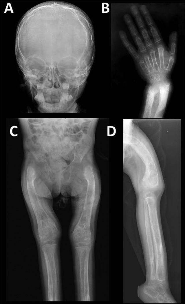
Figure 5. Radiographic Findings at Referral at 2.5 Years-of-Age.
A) The skull has a wide diploic space with coarse vertically oriented trabeculae and an indistinct thin outer table. The sagittal suture is closed.
B) Radiographs of the hand and wrist shows severe osteopenia. The short tubular bones are wide, especially the phalanges, and there are thin cortices. The radius and ulna show metaphyseal and metadiaphyseal sclerosis, loss of corticomedullary definition, thin cortices, distal flaring, and slight fraying of the provisional zones of calcification. Soft tissue swelling is apparent.
C) The femurs and tibias are osteopenic, wide, and have irregular cortical thickening, indistinct corticomedullary junctions, focal areas of sclerosis and lucencies, marked coxa vara, failure of modeling (especially in the distal femora), and lateral femoral bowing. There is mild acetabular protrusion.
D) This leg and distal thigh demonstrate tibial and femoral bowing, wide medullary cavities with indistinct margins, irregular cortical thickening, and interference with modeling. Coarse and thick trabeculae and focal areas of sclerosis and lucencies are present.

