


Fig 1.
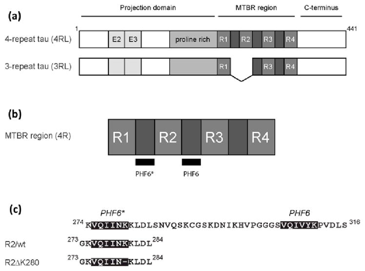
Tau isoforms, location of PHF6* and PHF6, and constructs used in this study. (a) The longest tau isoforms contain either three or four imperfect repeats in the microtubule-binding repeat region (MTBR). The amino terminal half of each protein contains two alternatively spliced exons (E2 and E3) and a proline rich region. (b) The MTBR region of 4-repeat tau, showing the locations of PHF6* and PHF6.(c) A closer view of PHF6*, PHF6, and the intervening sequence. The sequence of the two tau peptides used in this study (R2/wt and R2/ΔK280) are shown.