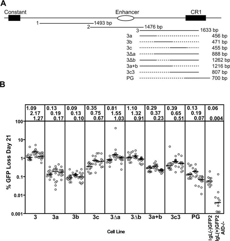
Figure 4. Deletion analysis of fragment 3.

(A) Schematic diagram of the location of fragments 1, 2, and 3 in the region 3′ of the chicken IgL locus. The structures and lengths of the various fragment 3 deletion mutants analyzed are indicated, with dotted lines indicating missing sequence. (B) Fluctuation analysis of GFP loss in subclones, with data depicted as in Fig. 2D.
