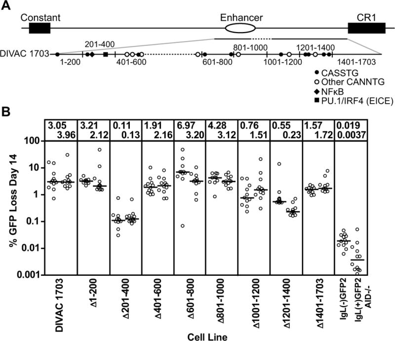
Figure 5. Deletion analysisof DIVAC 1703.

(A) Schematic diagram of the location of the DIVAC 1703 fragment in the region 3′ of the IgL locus. The DIVAC 1703 fragment is enlarged below to illustrate the end points of deletions (black vertical lines) and relevant DNA motifs (see key; S=G or C). Dotted lines indicate missing sequence. (B) Fluctuation analysis of GFP loss in subclones with data depicted as in Fig. 2D. The cell lines and test fragments are named for the roughly 200 bp segment that was deleted from DIVAC 1703.
