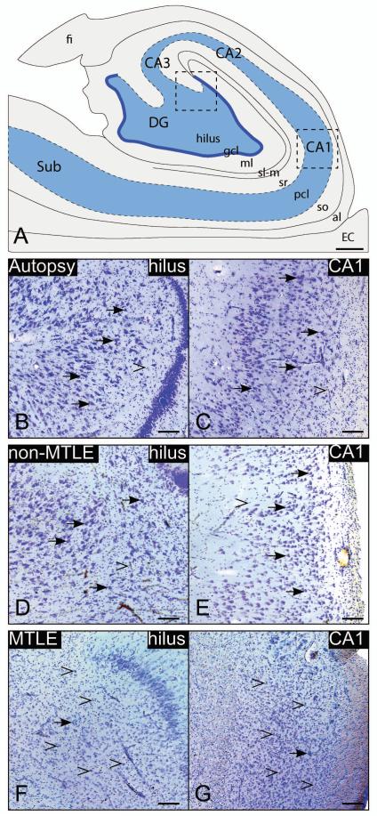
Fig. 1. Nissl-stained coronal sections from representative cases.
High-power images from the dentate hilus (B, D, F) and CA1 (C, E, G) of the hippocampal formation (A) are presented. Hippocampal formations from autopsy and non-MTLE subjects are characterized by abundant neurons (arrows) in the hilus (B, D) and CA1 (C, E). In contrast, there is marked neuronal loss in the hilus (F) and CA (G) in the hippocampal formation in MTLE subjects. The loss of neurons in these areas is accompanied by reactive gliosis (open arrowheads in F, G). Little or no reactive gliosis is present in autopsy (B, C) or non-MTLE (D, E). Abbreviations: al, alveus; EC, entorhinal cortex; CA1-3, cornu ammonis 1-3 of the hippocampus; DG, dentate gyrus; fi, fimbria; gcl, granular cell layer; ml, molecular layer of the DG; sl-m, stratum lacunosum-moleculare; so, stratum oriens; pcl, stratum pyramidale; sr, stratum radiatum; SUB, subiculum. Bars: A, 1 mm: B-G, 200 um.

