Figure 2.
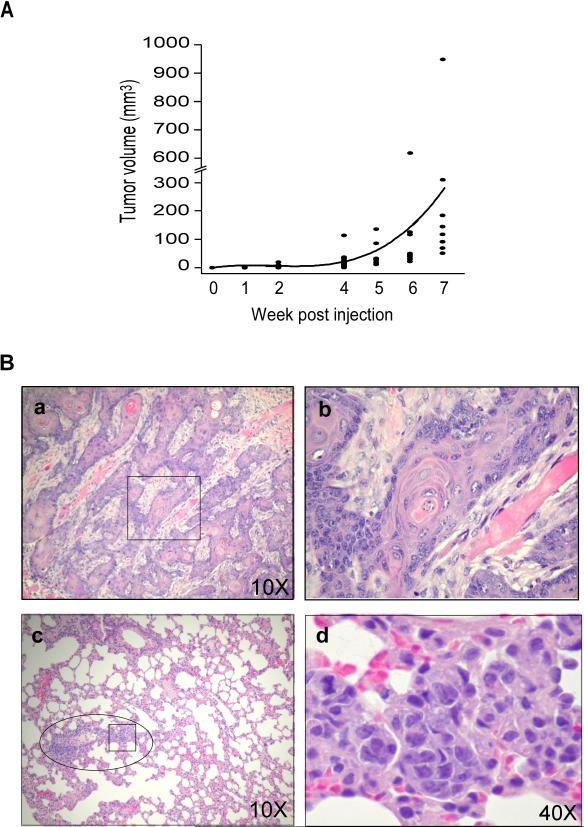
Time course of tumor development and tumor size and histology of SCCs from athymic nude mice injected s.c. with ONSC cells. (A) Time course of ONSC tumor development. ONSC cells (2×10⁶) were injected into the lower dorsal regions of female nude mice. Tumors were measured each week (except wk 3) for 7 wk. The curved line represents mean tumor volumes at each week (n=8/group/wk). (B) Histology of SCC in athymic nude mice injected with ONSC cells. Athymic nude mice were injected s.c. with ONSC cells. (a and b) Well differentiated SCC at 1 wk after injection of ONSC cells. (c and d) SCC metastasized to the lung of athymic nude mice. (a and c), magnification 10x; (b d), magnification 40x. □, magnified regions in b and d; ○, region with SCC in the lung.

