
Figure 2. Chronic chromium exposure represses E-cadherin and enhances vimentin protein expression.

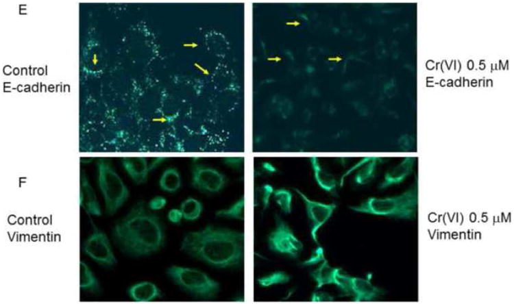
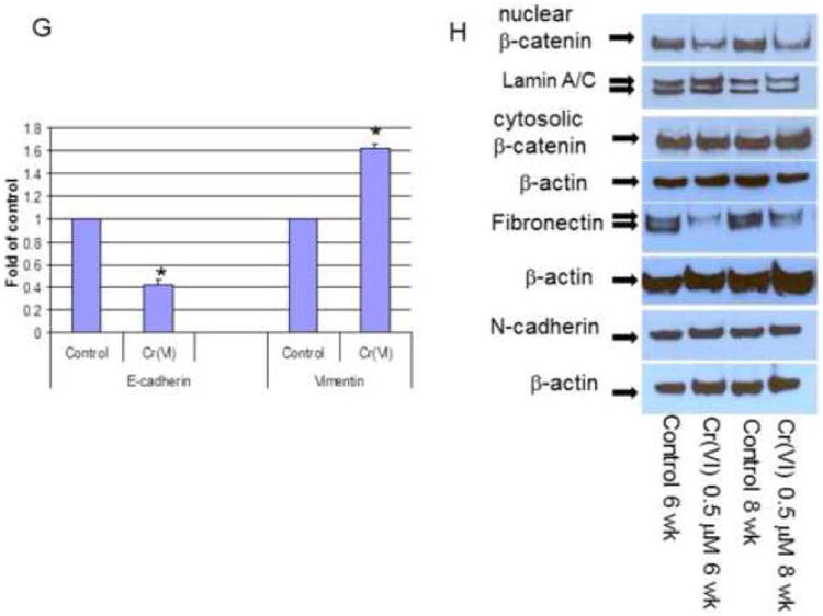
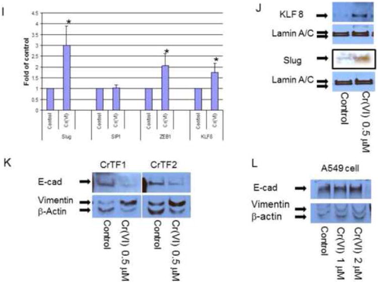
To evaluate E-cadherin related protein expression, BEAS-2B cells were treated with or without Cr(VI) at 0.5 μM for 3-10 weeks (A-J). Cytosolic and nuclear proteins were extracted and separated on 10% SDS-polyacrylamide gel to detect E-cadherin (E-cad), vimentin, Snail, Twist, β-catenin, N-cadherin, fibronectin, krupple-like factor 8 (KLF 8) and Slug protein expression (A-D, H, J). Blots are representative of three separate experiments with similar results, arrows indicate specific band. For immunofluorescence staining of E-cadherin and vimentin, cells were seeded at 1 × 10⁴ in 8 well chamber, fixed with 1% formalin, washed and stained with E-cadherin and vimentin antibody overnight at 4°C. After washing and secondary antibody conjugation, cells were visualized with immunofluorescence microscope (E, F). For gene expression assay, total RNA was collected from above cells and real-time PCR was performed to detected E-cadherin, vimentin, Slug, SIP1, ZEB1 and KLF8 mRNA expression (G, I). Data are mean±SEM from three separate experiments, *P<0.01 when compared with controls. To determine E-cadherin and vimentin protein expression in tumor cells, CrTF1, CrTF2 and A549 cells were treated with or without Cr(VI) at 0.5 μM for 3 weeks, cell lysates were extracted to detect E-cadherin and vimentin protein expression (K, L) as described above.