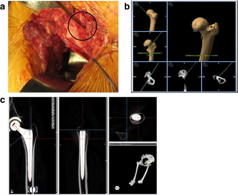
Fig. 2.
a Postoperative CT data was transferred to the planning module and was reconstructed to axial, frontal, and sagittal planes. The computer-aided design model of the femoral implant was superimposed. b Verification point of the anterior side of the femur. c The position of the pointer in the dialogue views shows a distance of 1.4 mm from the bone

