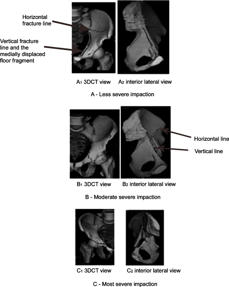
Fig. 4.
a Less severe C fracture, 3D AP image (A1) shows left central hip dislocation. Interior lateral (IL) view (A2) shows a triangular shaped fragment of broken Q surface or acetabular floor. b Moderate severe C fracture (B1) 3D AP image and IL view (B2) show a moderate severe injury by the extension of the primary vertical fracture line. c Most severe C fracture (C1) 3D AP image and IL view (C2) show severe impaction by the aggressive extension of primary and secondary fracture lines breaking through columns

