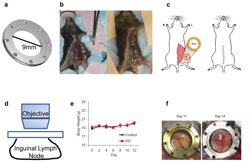
Figure 4. Development of the lymph node window chamber technique.
a) Schematic of the titanium lymph node window chamber. b) Representative picture of a mouse after implantation of the LNIWC over the mouse ILN (left). The surgical clips are used to close the skin until the next imaging session (right). c) Schematic of the placement of the LNIWC after surgical implantation (left) over the ILN, and a schematic of the mouse after closing the skin using surgical clips (right). d) Schematic of IVM being performed using the LNIWC. e) Body weight measurements of C57BL/6 mice either carrying the LNIWC (WC) or not (control). Symbols represent the means of body weight. N=3 mice per group in a single independent experiment. Bars: S.E.M. f) Representative pictures of tumor growth in the LNIWC.

