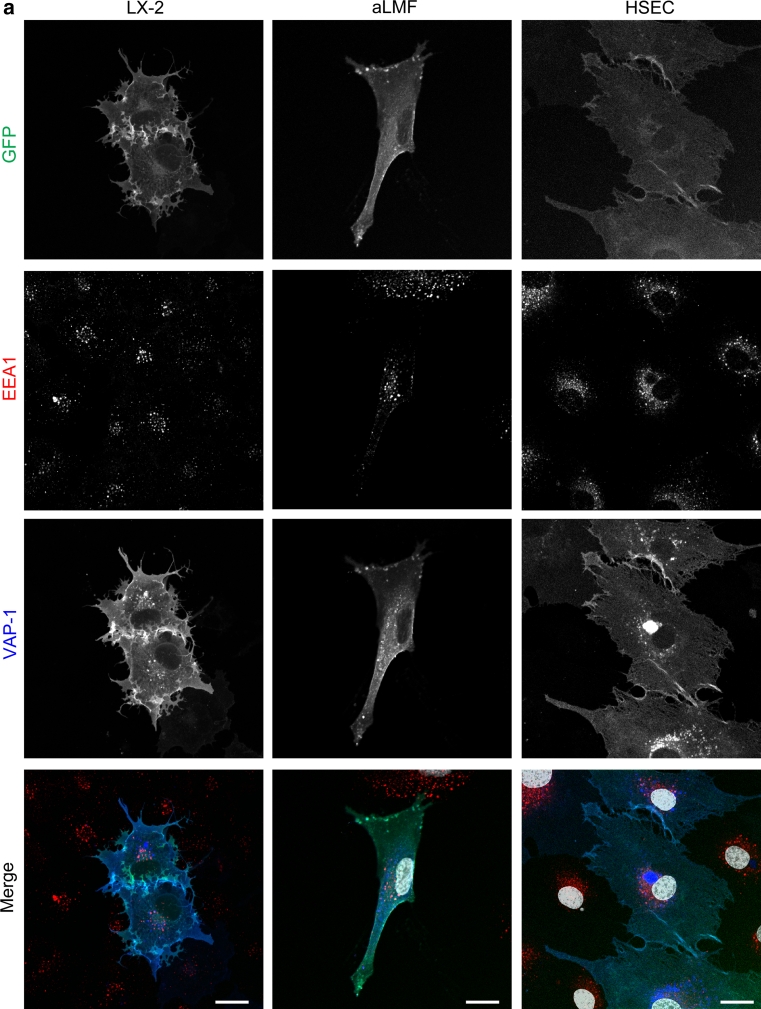
Fig. 3.
GFP-wtVAP-1 localizes to endosomes and other cellular vesicles. a Multicolour confocal microscopy indicated that GFP-wtVAP-1 co-localized with the early endosomal marker EEA-1. Merged images: GFP green; EEA-1 red; VAP-1 blue; nuclei white. b The distribution of GFP and anti-VAP-1 antibody fluorescence in live and fixed cells revealed the presence of intracellular GFPlowVAP-1high vesicles in fixed, but not live cells. Merged images: GFP green; VAP-1 red; nuclei blue. Scale bar 20 μm


