
FIGURE 1.
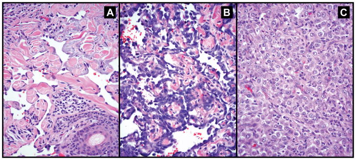
Representative images of the histologic spectrum of angiosarcoma reviewed in this series. (a) vasoformative angiosarcoma showing well-formed irregular anastomosing vascular channels dissecting dermal collagen; the endothelial cells show mild cytologic atypia and lack mitoses; (b) vasoformative angiosarcoma with high grade nuclear features and (c) epithelioid angiosarcoma composed of solid sheet of polygonal cells resembling carcinoma; vascular channels are not seen.