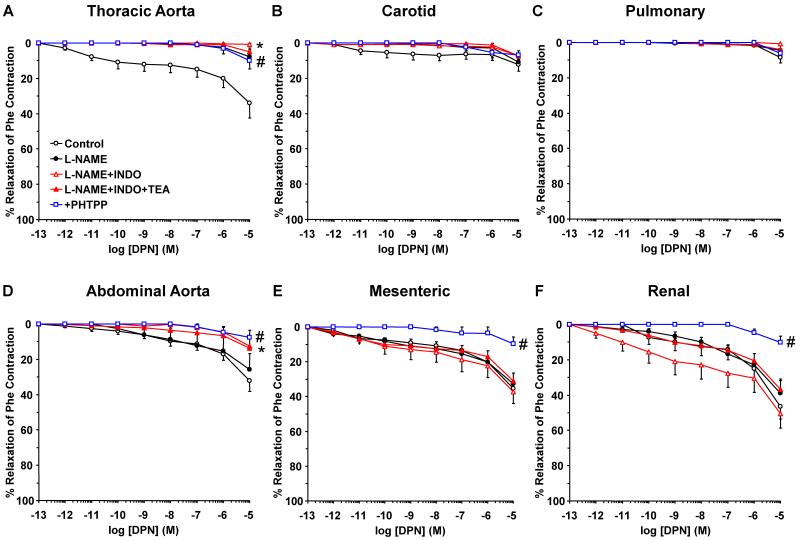
Fig. 8.
Contribution of NO, PGI2, and hyperpolarization factor to ERβ-mediated relaxation of cephalic, thoracic and abdominal arteries of female rat. Endothelium-intact segments of thoracic aorta (A), carotid (B) pulmonary (C) abdominal aorta (D), mesenteric (E) and renal artery (F) were either nontreated or pretreated for 15 min with L-NAME (3×10⁻⁴ M), L-NAME+indomethacin (INDO, 10−5 M), or L-NAME+INDO+TEA (30 mM). The vessels were precontracted with submaximal concentration of Phe then increasing concentrations (10−12 to 10−5 M) of DPN (ERβ agonist) were added and the % relaxation of Phe contraction was measured. The specificity of the relaxation effects of DPN were tested in blood vessels pretreated with the ERβ antagonist PHTPP (10−5 M). Data represent means±SEM, n= 8 to 10. * Maximal relaxation is significantly different (p<0.05) from corresponding measurements in control nontreated vessels. # Measurements in vessels treated with the ERβ antagonist PHTPP are significantly different (p<0.05) from corresponding measurements in control nontreated vessels.

