Figure 6.

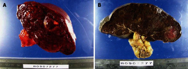
Macroscopic findings of the spleen. A: The raw specimen. Multiple nodules with different colors from normal splenic tissue; B: The specimen after formalin fixation. The arrows show the tumor.

Macroscopic findings of the spleen. A: The raw specimen. Multiple nodules with different colors from normal splenic tissue; B: The specimen after formalin fixation. The arrows show the tumor.