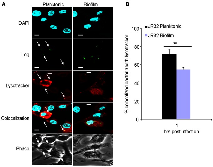
Figure 4.
Vacuoles harboring biofilm-derived L. pneumophila bacteria significantly evade fusion with lysosomes. (A) Representative images of WT BMDMs infected for 1h with JR32 planktonic or biofilm. Nuclei are stained blue with DAPI and L. pneumophila stained green with L. pneumophila-specific antibody. Lyso-tracker red was used to stain acidified lysosomes. White arrows indicate L. pneumophila colocalization with lysotracker. (B) Percent colocalization of L. pneumophila with lysotracker. Images were captured with the 60× objective and magnified 3×, scale bar = 10 μm. Data are presented as means ± SD of three independent experiments each performed in triplicates. Asterisks indicate significant difference (**P < 0.01).

