Fig. 1.
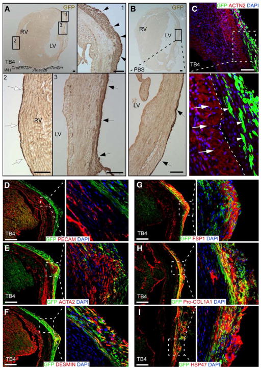
Reactivated epicardial layer and cell fate after myocardial infarction followed by thymosin β4 (TB4) treatment. After myocardial infarction followed by TB4 or PBS treatment, Wt1CreERT2/+;Rosa26mTmG/+ hearts were analyzed by immunochemistry. (A, B) Immunostaining for GFP in TB4-treated (A) or PBS-treated (B) heart. GFP marks EPDCs. The GFP+ epicardial layer covering the heart was thicker in the peri-infarct (arrowheads, inset 1) and infarct (arrows, inset 3) regions with TB4 treatment. The epicardial layer remained thin in the remote (right ventricular) region (inset 2). LV, left ventricle; RV, right ventricle; bar=100 μm. (C) EPDCs (white arrowheads) did not migrate into myocardium or express cardiomyocyte markers in Wt1CreERT2/+;Rosa26mTmG/+ hearts that underwent experimental infarction followed by TB4 treatment. White arrows indicate cardiomyocytes and white arrowheads indicate EPDCs. White dotted line indicates the border between myocardium and the epicardial layer. Bar=100 μm. (D–I) In infarcted, TB4-treated heart, EPDCs (GFP+) did not differentiate into coronary endothelial cells, marked by PECAM. A subset of cells expressed smooth muscle actin (ACTA2) or DESMIN, markers of myofibroblast and smooth muscle cells. Many GFP+ cells also co-expressed fibroblast markers FSP1, Pro-Collagen I and HSP47. Bar=200 μm.

