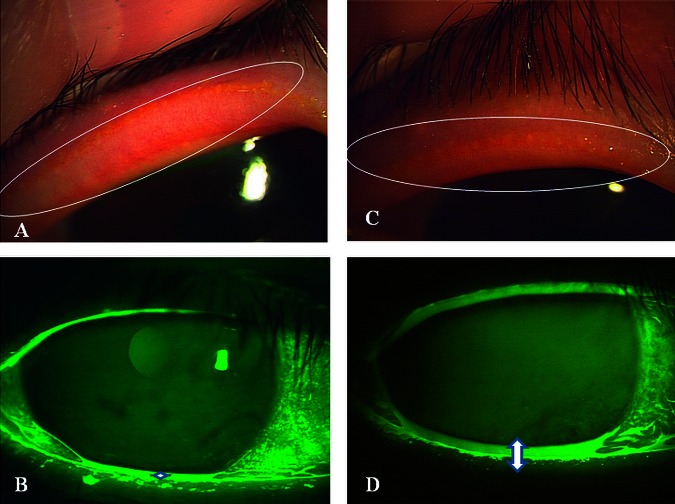
Figure 2.
Representative case of a 66-year-old woman with obstructive meibomian gland dysfunction (case #5). (A) Lid margin vascularity and pluggings of the orifices were observed before therapy. (B) Tear meniscus height was very low (0.1 mm). (C) Lid margin vascularity and pluggings of the orifices decreased after 8 months of therapy. (D) Tear meniscus height was increased (0.3 mm).

