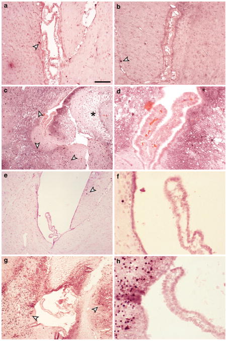
Fig. 5.
Caspase-3 immunoreactivity after systemic LPS and hypoxia-ischemia (HI). In control tissue (a) as well as 24 h after 1 mg/kg LPS (b), occasional cells were positive for caspase-3 (arrowheads). At 24 h after HI a great number of caspase-3 positive cells were found in hippocampal and thalamic regions surrounding the lateral ventricle in the ipsilateral hemisphere, however, no positive cells were found in the choroid plexus. Lesions were also beginning to appear in tissue at this time (asterix in C). d is higher power micrograph from C showing that none of the choroidal epithelial cells were caspase-3 positive. In HI20 animals, the brain region around the lateral ventricles showed few caspase-3 positive cells (e) with no positive cells in the plexus (f) and in LPS/HI animals extensive caspase-3 labeling was present around ventricles (g) yet no positive cells in plexus (h). Note that all sections were counterstained with acid fuchsin and that no apparent abnormalities were found in choroidal epithelial cells after treatments. This indicates no obvious damage in the choroidal barrier forming cells after either LPS or HI. Scale bar represents 100 μm in A&B, 200 μm in C, E, G and 50 μm in D, F, H

