
Fig. 1.
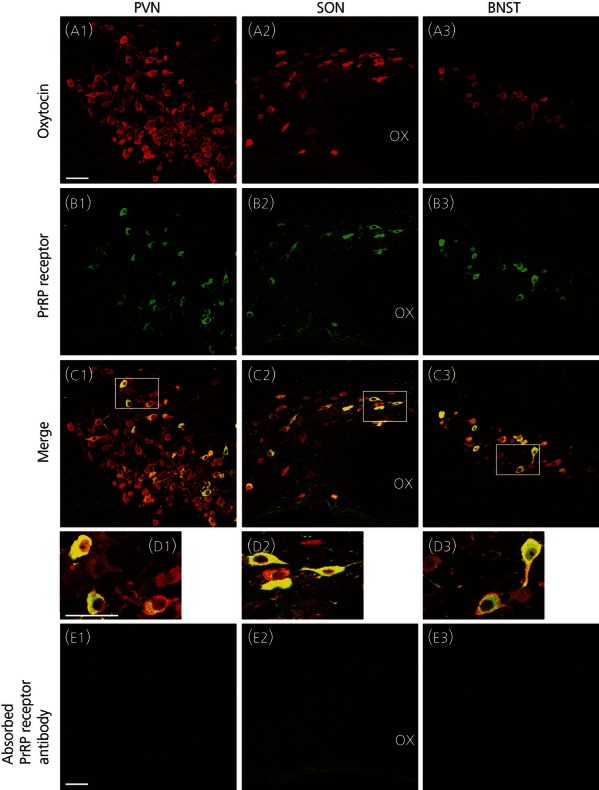
Expression of prolactin-releasing peptide (PrRP) receptors in oxytocin-immunoreactive (-IR) neurones in the hypothalamus or bed nucleus of the stria terminalis (BNST) in rats. Immunoreactivity for oxytocin (red fluorescence, a) or PrRP receptors (green fluorescence, b) is shown. Double-labelled cells are shown in yellow (c, d). Areas enclosed by white rectangles in the merged images (c) are shown at a higher magnification (d). Immunoreactive PrRP receptors were observed in oxytocin-IR cells of the paraventricular nucleus (PVN, a1, b1, c1, d1), supraoptic nucleus (SON, a2, b2, c2, d2) and BNST (a3, b3, c3, d3) in rats. All labellings for PrRP receptors were prevented by pre-absorption of anti-PrRP receptor antibody with an excess of the synthetic antigen peptide (e). The third cereberoventricle (not shown) is located on the right side of each picture. OX, optic chiasma. Magnifications in (a–c) and (e) are equivalent. Scale bars = 50 μm.