
Fig. 2.
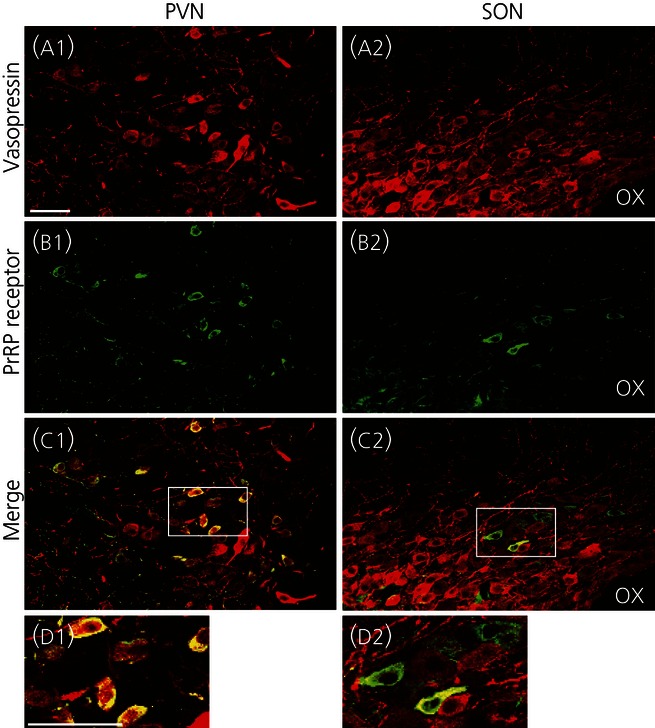
Expression of prolactin-releasing peptide (PrRP) receptors in vasopressin-immunoreactive (-IR) neurones in the hypothalamus of rats. Immunoreactivity for vasopressin (red fluorescence, a) or PrRP receptors (green fluorescence, b) is shown. Double-labelled cells are shown in yellow (c, d). Areas enclosed by white rectangles in the merged images (c) are shown at a higher magification (d). Immunoreactive PrRP receptors were observed in vasopressin-IR cells of the PVN and SON. OX, optic chiasma. Scale bars = 50 μm.