
Fig. 5.
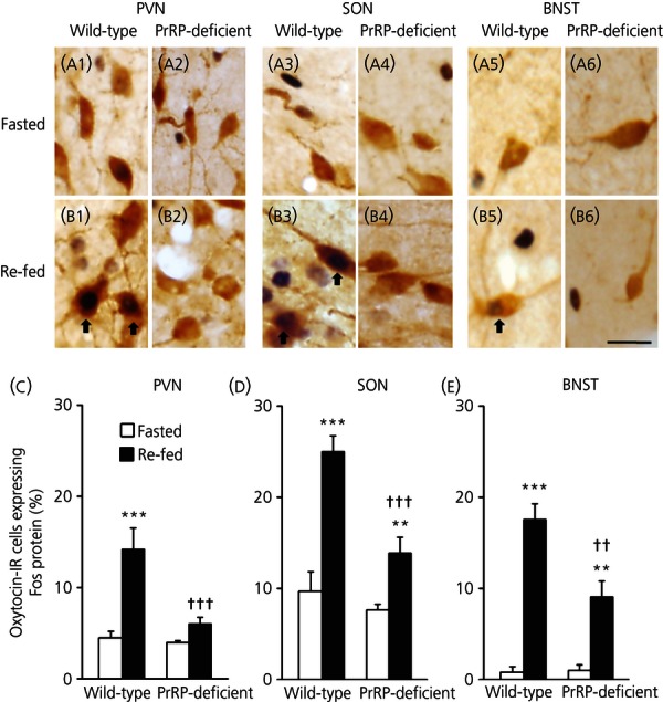
Expression of Fos protein in oxytocin-immunoreactive (-IR) neurones of the hypothalamus or bed nucleus of the stria terminalis (BNST) following re-feeding in prolactin-releasing peptide (PrRP)-deficient mice. Images of oxytocin-IR cells and Fos protein immunoreactivity in the paraventricular nucleus (PVN) (a1, a2, b1, b2), supraoptic nucleus (SON) (a3, a4, b3, b4) and BNST (a5, a6, b5, b6) of wild-type or PrRP-deficient mice in fasted or re-fed groups and the percentages of oxytocin-IR cells expressing Fos protein (c–e) are shown. The percentage of oxytocin-IR neurones expressing Fos protein following re-feeding was significantly decreased in the PVN, SON or BNST of PrRP-deficient mice compared to that in wild-type animals. Scale bar = 20 μm **P < 0.01 and ***P < 0.001 compared to fasted mice. ††P < 0.01 and †††P < 0.001 compared to corresponding groups of wild-type mice (n = 5–8). Arrows indicate double-labelled neurones.