
Fig. 6.
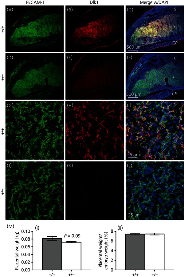
Embryonic development of the placenta in the delta-like 1 homologue (Dlk1)-null mutant. Sections from placentas of wild-type and Dlk1-null mutant E18.5 embryos stained for platelet endothelial cell adhesion molecule-1 (PECAM-1) (green), Dlk1 (red) and 4′,6-diamidino-2-phenylindole (DAPI) (blue). Expression of Dlk1 in the placenta is predominantly in the labyrinth, where PECAM-1 is expressed in a large number of foetal blood vessels (a–c); although Dlk1 expression is lacking in the mutant embryo, the foetal labyrinth appears to have formed normally (d–f). S, spongiotrophoblast; L, labyrinth; CP, chorionic plate. Low magnification scale bar = 500 μm. Dlk1 colocalises with many of the PECAM-1-expressing endothelial cells in the wild-type embryo (g–i). The expression of PECAM-1 is not affected by the loss of Dlk1 in the mutant embryo (j–l). High magnification scale bar = 25 μm. (m, i) Placental weights of Dlk1-null mutants are slightly, but not significantly (Dlk1-null versus wild-type littermates), reduced at E18.5. (m, ii) Placental weights expressed as a percentage of the embryo weight shows no difference between the wild-type and Dlk1-null embryos, showing that placental weights are reduced to the same extent as the embryo weights. Data are shown as the mean ± SEM of three to five embryos.