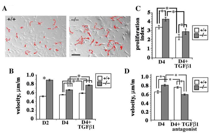
Figure 2. Primary sdc-1 null fibroblasts migrate faster than wt cells.
A. Time lapse studies of wt and sdc-1 null fibroblasts. The tracks (indicated in red) for the null fibroblasts were longer than those for the wt fibroblasts suggesting that the null cells were migrating faster. Bar = 10 μm B. The sdc-1 null cells migrate faster at days 2 and 4 as well as after TGFβ1 treatment; wt cells had similar velocities regardless of time point or TGFβ1 treatment. C. Proliferation indices showed that at day 4, the sdc-1 null fibroblasts proliferated faster than wt cells. Both genotypes responded to addition of TGFβ1 by reducing their proliferation rate. D. Antagonizing TGFβ1 activity using a TGFβ1 function blocking antibody inhibited null cell migration but had the opposite affect on wt cells. Data in B-D indicate mean values +/− SEMs for a minimum of 60 individual cells for each variable investigated; asterisks show data that were significantly different with p < 0.05 as determined using the student’s t test.

