Figure 1.

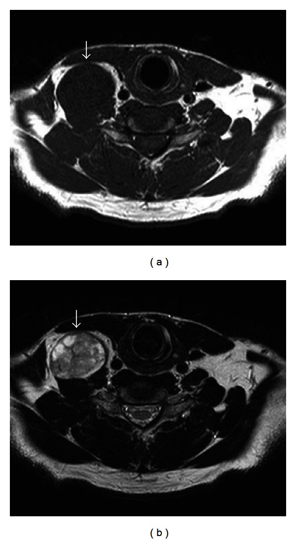
MRI findings for case 24. (a) Axial T1-weighted imaging showed a mass with signal hypointensity (arrow). (b) Axial T2-weighted imaging showed a mass with heterogeneous signal hyperintensity (arrow).

MRI findings for case 24. (a) Axial T1-weighted imaging showed a mass with signal hypointensity (arrow). (b) Axial T2-weighted imaging showed a mass with heterogeneous signal hyperintensity (arrow).