
Figure 4.
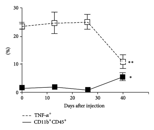
Frequency and functional changes in tumor-associated macrophages and microglia (TAMs) in ex vivo tumor specimens over the course of tumor development. While the percentage of TAMs (CD11b+CD45+ cells) was increased by the final time point (1.1% versus 5.6%, P = 0.017), their functional capacity was impaired as measured by TNF-α expression (25.2% versus 10.9%, P = 0.007). Thirteen mice were analyzed at 13 dpi (n = 4), 26 dpi (n = 4), and at 29 dpi upon exhibiting clinical tumor morbidity (n = 1) or at 40 dpi (n = 4). Differences between the means at each time point were tested using two-sided t-tests with unequal variances. *P < 0.05, **P < 0.01, ***P < 0.001. Published with permission from Kennedy et al. [21].